AI technology could help tackle leading cause of blindness
A global research team has developed Artificial Intelligence (AI) algorithms that will automate the screening process of remote eye tests for Diabetic Retinopathy (DR), a common complication of diabetes that can cause blindness if left untreated.
This new technology could prevent blindness, whilst making huge cost-savings particularly in less developed countries.
The AI models, developed by the team from Queen’s University Belfast, The Philippine Eye Research Institute and Joslin Diabetes Center of Harvard Medical School, have been developed using local eye images taken in the Philippines, but have been shown to match current existing commercial algorithms.
DR is the leading cause of blindness among the working age population worldwide, and with 537 million adults currently living with diabetes globally; predicted to increase to 643 million adults by 2030, the incidence of DR is also expected to rise substantially. There is therefore a need to increase diabetic eye screening efforts to prevent blindness among people with diabetes.
However, the introduction of widescale systematic DR screening programmes (DRSP) is not straightforward, especially in countries such as the Philippines which faces challenges such as low resources, high population, lack of healthcare infrastructure and geographic isolation- as the country is composed of 7641 islands. Currently, there is no national programme for DR screening in the country.
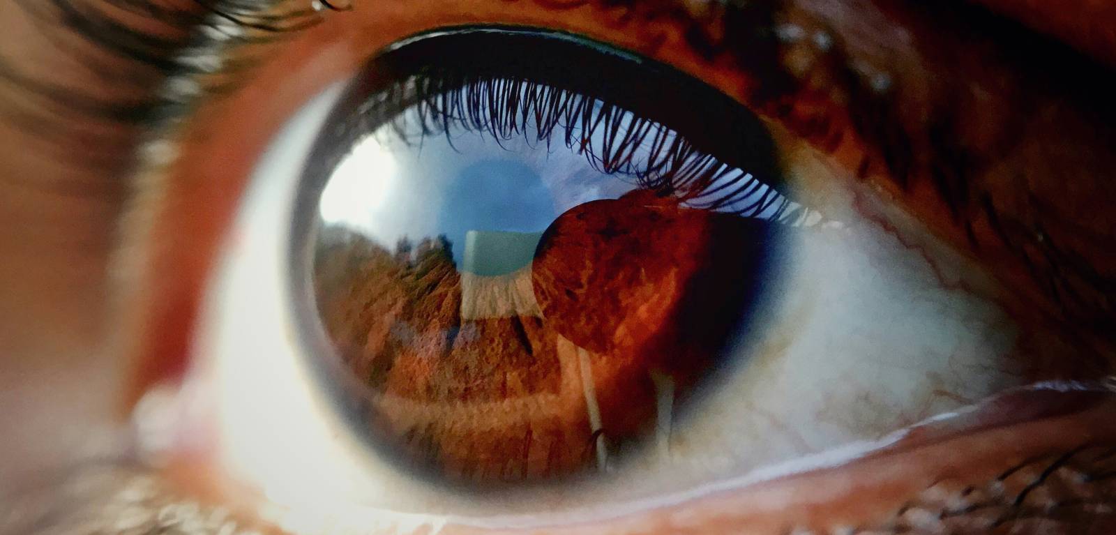
The use of digital technology and portable handheld retinal cameras operated by trained imagers are uniquely suited to addressing these issues. In this set-up, handheld retinal cameras, which are more portable and less expensive compared to traditional models of care, are used to take screening photographs of the patient’s retina (back part of the eye) to check for any signs of diabetic eye disease. The photographs are then sent electronically to a centralised reading centre where they are analysed by trained graders, and the results including follow-up details and recommendations are sent back to the patient and screening sites.
Even with using these handheld retinal imaging devices, the sheer volume of images that need to be evaluated by trained human graders in a timely manner is a huge challenge. Training and certification of human graders also takes time and requires considerable financial investment which can be prohibitive for many health systems. Automation of the image evaluation process via AI has the potential to increase throughput and also maintain cost-efficiency.
In this study the researchers created code-free automated machine learning models (AutoML) for DR screening using images from handheld retinal cameras. The development of these AI algorithms allows users to perform a specific task to address a critical clinical need. These locally developed AI algorithms may potentially allow more images to be assessed, more accurately and at less cost.
The combination of community-based DR screening and local point of care integration of AI has the potential to increase efficiency and lower the cost of screening for DR in settings such as the Philippines, where access to specialised care is difficult in remote, rural communities.
Professor Tunde Peto, Professor of Clinical Ophthalmology from the School of Medicine, Dentistry and Biomedical Sciences at Queen’s said: “AutoML allows the development of code-free algorithms at minimal cost by individuals without extensive background in computer programming language. As coding skills are not common among healthcare workers, the use of AutoML models for DR screening can potentially address disparities in the delivery of eye care to patients with diabetic eye disease by ensuring a more rapid assessment of retinal images at point of care with minimal cost, thereby ensuring prompt referrals and timely intervention for those patients who require more specialised eye care.
“Our research in the Philippines has shown that this is particularly useful in low-resource settings where there is a huge clinical burden of diabetes, a high cost of services and a lack of eye care human resources. This work can help clinicians and healthcare managers in the planning and scaling-up of DRSP operations, with the ultimate goal of saving sight in people with diabetes.”
Featured Expert
Media
Media inquiries to Jemma Coulter from Queen's Communications Office at jemma.coulter@qub.ac.uk.